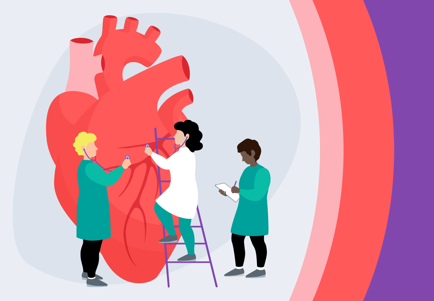
Cardiac Diagnostics & Imaging
Cardiac diagnostics and imaging help our cardiovascular experts
understand your heart and create the best treatment plan to optimize your heart health.
Using advanced imaging technology to provide a clear picture of your heart's function,
we offer a range of tests to diagnose and monitor heart conditions,
helping you get the peace of mind you deserve.
- Cardiac Diagnostics and Imaging Services
- About
- PeopleCare Stories
- Services
- Locations
About
Advanced Heart Technology & Expertise
While cardiologists may perform some tests in their office, they may refer you for more advanced imaging. We use advanced technology to make and analyze sophisticated images of the heart.
The cardiac imaging and diagnostics team supports your team of heart care specialists in understanding what is causing symptoms. Imaging is also often needed to monitor your heart to see if medicines and other treatments are working.
Services
Types of Diagnostics & Cardiac Imaging
Our cardiovascular experts use a combination of tests to make the most accurate diagnosis. Some cardiac diagnostic tests are performed in the office by your cardiologist or by a lab. To provide you with the most accurate diagnosis, your doctor may refer you to a specialized cardiac diagnostic center for these types of imaging and tests:
- Calcium CT scoring (heart screening)
- Electrocardiogram (EKG)
- Echocardiogram
- Transesophageal echocardiogram
- Cardiac monitors
- Stress tests and stress tests with imaging
- Cardiac CT & Cardiac CT with FFR (HeartFlow)
- Cardiac MRI
- Nuclear imaging/SPECT study
- Tilt table testing
- Cardiac catheterization
Calcium CT scoring
Calcium CT scoring is a non-invasive test that detects calcified plaque in the coronary arteries and rates a person’s risk level for heart attack or coronary artery disease. This test is performed through our Know Your Heart Screening program.
Electrocardiogram (EKG)
An electrocardiogram (EKG) is a painless test commonly used to monitor electrical signals in the heart. An EKG can help detect irregular heart rhythms, blocked or narrowed arteries or learn whether someone has had a heart attack. It can also indicate whether treatments are working. We typically conduct EKGs with a Holter Monitor, a portable device worn on the chest.
Echocardiogram
An echocardiogram—or echo—is a non-invasive ultrasound that sends high-frequency soundwaves to create images of the heart. It helps your doctors analyze both the heart structures and blood flow within. The purpose is to see if every part of the heart wall is helping the heart pump normally. Weakened areas may be a sign of coronary artery disease.
Transesophageal echocardiogram (TEE)
A transesophageal echocardiogram produces more detailed images of the heart with sound waves than a standard echocardiogram. An interventional cardiologist uses a small tool that makes sound waves—a transducer connected to a thin tube—and passes it through the mouth, down the throat and into the esophagus. This provides clearer images than a traditional echocardiogram because the sound waves are closer to the heart and don’t have to pass through skin, bone and muscle.
Cardiac monitors
A cardiac monitor is a specialized device that records the electrical activity of a person’s heart for a set amount of time. There are a few options including portable devices like the Holter monitor, which continuously records the heart’s rhythm for 24 to 48 hours. Other devices can be implanted or worn on the body to track heart activity for a longer period. For example, a loop recorder implanted underneath the skin can monitor a person’s heart for up to three years.
Stress tests
A stress test looks for signs that the heart is pumping blood efficiently or inefficiently. Also known as an exercise stress test, this evaluation is performed on a treadmill or stationary bike while monitoring a person’s blood pressure, breathing and heart rate. A stress test may be recommended if someone has symptoms of a heart condition or to see how well a heart treatment is working.
To learn more about the heart’s performance, a stress test can be combined with an echocardiogram or 3D imaging such as a cardiac CT, a cardiac MRI or nuclear imaging/SPECT study. These advanced imaging tests aid in more accurate diagnosis and procedure planning.
Cardiac CT
A cardiac computed tomography CT scan takes a specialized X-ray of the heart. The scan looks for plaque build-up, a substance made of fat and calcium that can narrow or close the arteries that supply blood to the heart. When a cardiac CT suggests that there may be a blockage, Wellstar physicians can analyze the fractional flow reserve (FFR) with leading-edge HeartFlow Analysis technology. This test details how each blockage impacts blood flow to the heart.
Cardiac MRI
A cardiac MRI is a type of imaging that uses large magnets and radio frequencies to see the heart valves and major vessels. It can help detect coronary artery disease and how much damage it has caused, assess heart problems that have been present since birth and identify tumors. It is often used to prepare for heart procedures and surgery.
Tilt table testing
A tilt table test evaluates the cause of repeated, unexplained moments of dizziness and fainting. This test can help trigger the symptoms while also monitoring heart rate and blood pressure.
Cardiac catheterization
A cardiac catheterization is a test and treatment for heart blockages. Guided by imaging, an interventional cardiologist inserts a catheter into an artery in the groin or arm to travel to the heart to look for blockages in the coronary arteries. We inject dye through the catheter to help blood vessels appear on the image. If a blockage requires treatment, a balloon can be inserted through the catheter and inflated to improve the blood flow. A mesh tube (stent) keeps the dilated artery open.
PeopleCare Stories