CT Enterography
Study Description
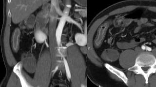
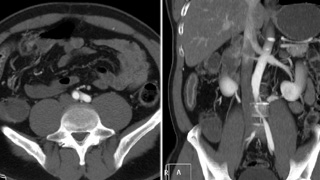
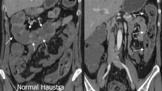
CT Enterography is an examination targeted to the small bowel. It replaces other small bowel studies such as a small bowel follow through under fluoroscopy. It is sensitive for detection of inflammatory bowel disease or small bowel masses. Outside of the bowel, it provides that same information as a conventional contrast enhanced CT. It is excellent for depiction of extraluminal pathology including abscesses and free intraperitoneal air from bowel perforation.
It is not a replacement for upper or lower endoscopy. Bowel motility is not evaluated, and MR Enterography may be a more suitable exam in some patients.
Patient Selection and Indications
Exam is optimal for patients with suspected inflammatory bowel disease or small bowel masses, especially as a first test and as a substitute for a conventional CT. It may permit evaluate gastric and colonic mucosa, but is not as sensitive as endoscopy or virtual colonoscopy for this purpose.
Serial scans increase radiation dose—a potential issue in young patients with Crohn’s disease. MR Enterography may be a more appropriate exam for serial follow up.
Use CT Enterography instead of MRE for:
- First presentation or baseline exam, particularly in a patient with prior surgery where anatomy has not been defined.
- Suspected abscess or in the septic patient
- The ER patient or in the context of suspected surgical emergency such as bowel perforation
Use MR Enterography instead of CTE for:
- Routine follow-up exam to spare radiation dose. This is most important in young patients and in particular in females of child bearing age.
- Known Chrohn’s disease, especially when there is a question of disease activity causing nonspecific symptoms such as pain.
- Assessment of IBD therapeutic efficacy. Biologic agents such as anti-TNF antibodies are quite expensive. Immunomodulatory or immunosuppresive medication may have significant side effect profiles. It is important to determine if the patient is responding as soon as possible to permit optimal therapy.
- Evaluation of intermittent partial bowel obstruction. Test is most useful if performed when the patient is symptomatic.
Patient Preparation
Please specify “CT Enterography Abdomen and Pelvis” when prescribing. Patients do not receive standard positive (barium) oral contrast, but instead receive a negative oral contrast agent at the time of the exam. This negative contrast agent distends the bowel and permits detection of mucosal enhancement.
Patient should be NPO for 4 hours prior to the procedure.
These exams require IV contrast to evaluate the mucosa of the stomach, small bowel and colon. Standard precautions with regard to IV iodinated contrast are necessary. If iodinated contrast is contraindicated, there is no role for CTE. MR enterography, either without or preferably with gadolinium, may be considered as an alternative.
Reporting and Outcomes
CTE is excellent at depicting early changes of IBD and documenting complications such as abscess or fistula. As the abdomen and pelvis are both imaged in their entirety during a CTE, unrelated pathology in other organs may often be detected.
Every CT enterography exam is reviewed by a board certified radiologist with subspecialty training in advanced body imaging. A comprehensive report detailing findings in the abdomen, pelvis, and in the bowel is provided to the referring clinician. If urgent findings are encountered, these are relayed in real time by telephone. A copy of the examination and report is furnished to the patient upon request. The exam may also be reviewed directly with the radiologist in the radiology department at a Wellstar facility.
